Ultrasound in CMIB is a safe, fast and accurate imaging method used to evaluate internal organs, vessels and tissues. Radiation-free and completely painless, it allows early diagnosis of conditions and monitoring of treatments, being carried out by specialized imaging doctors with modern equipment.
Ultrasound is a non-invasive imaging investigation that uses ultrasound to obtain real-time images of internal organs. Sound-wavas emituje na ultrasonovska probe v a.
This method is one of the safest and most accurate forms of first-aid diagnosis. Ultrasound is used in almost all medical specialties — from internal medicine and cardiology, to gynecology, urology, endocrinology or pediatrics — because of its non-irradiating and affordable nature.
Ultrasound is recommended when the patient experiences symptoms such as abdominal pain, persistent bloating, urinary changes, swelling, palpable nodules or abnormal sensations in certain areas of the body. It is also indicated in case of trauma, suspicion of inflammation or infection, and in monitoring pregnancy.
Ultrasound is used frequently and for preventive purposes, as part of regular medical check-ups. Routine ultrasonizations can identify benign lesions, early changes in internal organs, or formations that can be effectively treated if discovered in time.
In patients undergoing treatment for liver, kidney, thyroid or vascular diseases, ultrasound allows monitoring the course of the disease and checking the effectiveness of therapy. It is also used for postoperative control or after interventional procedures.
Ultrasound is based on the use of high-frequency ultrasound, emitted by a special probe connected to an ultrasound machine. Testi velove sono se pokazati na tevne i organa, se reproducirati v forma immagini dynamici, visibili na monitora.
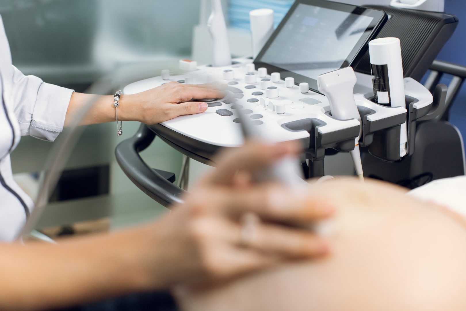
The patient is placed on the examination bed, depending on the area under investigation. The doctor applies a transparent gel to the skin, to facilitate the transmission of sound waves. Probe na, a. The examination generally takes between 10 and 30 minutes and is completely painless.
The results are immediately interpreted by the imaging doctor. In some cases, images may be archived for comparison with future examinations. Ultrasound report is provided to the patient on the spot, containing the clinical conclusions and possible recommendations for further investigation.
It examines the organs of the abdominal cavity — liver, gallbladder, pancreas, spleen and kidneys. It is used to diagnose liver, biliary, kidney or pancreatic diseases.
Aims to examine organs in the pelvis — bladder, uterus, ovaries in women and prostate in men. It is useful for detecting cysts, uterine fibroids, inflammations or prostate adenoma.
It allows to assess the size and structure of the thyroid gland, detect nodules, inflammation (thyroiditis) or other changes in the thyroid parenchyma.
It is an essential method of detection and monitoring of breast lesions, recommended in addition to mammography or as a periodic screening, especially in young women.
It uses the Doppler effect to analyze blood flow in arteries and veins, identifying possible blockages, thromboses or circulatory failure.
It visualizes tendons, ligaments, muscles and joints, being used to diagnose injuries, inflammation or degenerative diseases.
Ultrasound can identify conditions such as liver steatosis, biliary lithiasis, liver cysts, inflammation of the pancreas, or abdominal tumors. It is a first-line investigation for chronic abdominal pain.
It allows detecting kidney stones, urinary infections, congenital malformations or urinary tract obstructions. It is also useful in monitoring transplanted kidneys.
Ultrasound thyroiditis detecta nodules, inflammaciones y modificaciones structurales que puede influenciar a função do thyroid gland, contribuir a la diagnosia correttamente de endocrinosos.
Doppler ultrasonography detects circulatory problems such as deep vein thrombosis, atherosclerosis or chronic venous insufficiency, being essential for preventing vascular complications.
Preparation for ultrasound depends on the type of investigation. For abdominal ultrasound, fasting 4—6 hours beforehand is recommended to reduce intestinal gases. Para ultrasúscula películum, a volta é necessário, se el patiente deve consumo fluidos antes de examinação.
In the case of vascular or musculoskeletal ultrasound, no special preparation is required. The patient is advised only to wear comfortable clothes and avoid applying creams to the examined area. The doctor gives clear instructions for each type of ultrasound individually.
After performing the ultrasound, the patient can resume normal activities immediately. The investigation does not require a recovery period and has no side effects.
Resultados s, a. Depending on the conclusions, further treatments, analyzes or specialist consultations may be recommended.
Ultrasound is completely safe, without radiation exposure and no discomfort. It can be repeated at any time, including in pregnant women and children, without risk.
Resultados se obtenido en el spoto, y los imagens proporciona alta precisión. Modern equipment allows to detect minor changes in organs and monitor their evolution over time.
Par, ultrasona a.
Take care of your health and do not postpone an essential check-up! Schedule an appointment now for a general ultrasound and receive a complete and accurate assessment of your health condition. Our team of imaging doctors is ready to help you with professionalism and care.

