Endocrine ultrasound at CMIB is a safe and non-invasive imaging investigation, used for the analysis of the thyroid, parathyroid and adrenal glands. It allows early detection of nodules, inflammations and tumors, providing accurate and immediate results. The examination is quick, painless and carried out by doctors specializing in endocrinology and imaging.
Endocrine ultrasound is a non-invasive imaging exploration method that uses ultrasound to analyze the structure of the endocrine glands. It provides detailed images of the thyroid gland, parathyroid, adrenal glands, and in some cases, the pituitary gland, helping to diagnose and monitor endocrine disorders.
This investigation has a crucial role in detecting and evaluating structural changes in the endocrine glands, such as nodules, cysts, inflammation or tumors. Endocrine ultrasound does not involve radiation and can be performed whenever necessary, including in children, pregnant women and patients undergoing hormonal treatment.
The endocrinologist or family doctor may recommend an endocrine ultrasound when the patient experiences symptoms such as persistent fatigue, weight variations, palpitations, intolerance to cold or heat, swelling in the neck, menstrual disorders or difficulty swallowing. These signs may indicate thyroid disorders or other hormonal imbalances.
Endocrine ultrasound is used to follow up patients diagnosed with conditions such as autoimmune thyroiditis (Hashimoto), hyperthyroidism, hypothyroidism, or thyroid nodules. It is also useful in the evaluation of the parathyroid glands in case of suspicion of hyperparathyroidism, and in the analysis of the adrenal glands in complex hormonal disorders.
In people with a family history of endocrine diseases or exposure to risk factors (such as radiation to the neck), endocrine ultrasound is recommended as a preventive measure. Regular examinations can detect early changes that can be effectively treated early.
Endocrine ultrasound uses high-frequency ultrasonic waves emitted by a probe connected to a specialized apparatus. These waves are reflected by the tissues of the endocrine glands and transformed into detailed images displayed on the monitor.
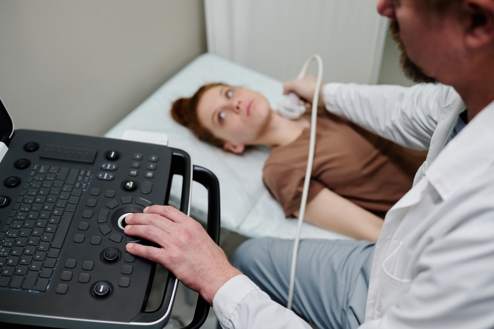
The patient is comfortably seated on the bed, with the neck slightly stretched in the case of thyroid examination. The doctor applies a conductive gel to the skin, then places the ultrasound probe in the area under investigation. The examination is completely painless, takes about 10—20 minutes and does not require anesthesia or hospitalization.
The images obtained are interpreted in real time by the specialist doctor, who gives a detailed report. It includes information about the size, shape, echogenicity and vascularization of the glands, as well as the presence of nodules or other changes. Depending on the result, hormonal analyzes, thyroid puncture, or other complementary investigations may be recommended.
It is the most common form of endocrine ultrasound. Evaluate the thyroid gland, identifying nodules, cysts, inflammation (thyroiditis), volume increases (goiter), or changes in structure. It is an essential investigation in the diagnosis of thyroid disorders.
It is carried out to identify and evaluate the parathyroid glands, located behind the thyroid. It is recommended in cases of hypercalcemia or suspicion of primary or secondary hyperparathyroidism.
It allows to visualize the adrenal glands, located above the kidneys, and is used to identify adrenal formations (adenoma, cyst, tumors). It is an important investigation in the evaluation of hormonal disorders and treatment-resistant blood pressure.
Ultrasound determines the presence, size and structure of thyroid nodules, allowing to differentiate solid ones from cystic ones. It also assesses the expansion of the goiter and the impact on neighboring organs.
It allows to identify inflammations of the thyroid gland, such as Hashimoto's thyroiditis or subacute thyroiditis, by the characteristic changes in the texture of the thyroid tissue.
Ultrasound detects benign or malignant tumor formations in the thyroid, parathyroid or adrenal glands, providing important information about size, vascularization and contour.
The investigation can highlight parathyroid adenomas or adrenal formations that influence hormonal secretion, being useful in evaluating hyperparathyroidism or Cushing's syndrome.
For ultrasound of the thyroid and parathyroid glands, no special preparation is required. The patient can eat and drink normally before the investigation. In the case of adrenal ultrasound, the doctor may recommend a few hours of fasting before the examination, for a clearer view of the abdominal organs.
It is important that the patient brings the results of previous ultrasounds and recent hormonal analyzes, in order to allow the doctor a comparative assessment. No contrast substance is administered and there are no restrictions after the examination.
Endocrine ultrasound does not imply a recovery period or restrictions. The patient can resume daily activities immediately.
The result of the ultrasound is provided on the spot, and the doctor explains the meaning of the images, describes any changes identified and recommends, if necessary, additional endocrinological consultation, blood tests or specific laboratory investigations.
Endocrine ultrasound is completely safe, without radiation, painless and without side effects. It can be repeated at short intervals, depending on the doctor's recommendation.
This investigation allows early detection of structural changes in the endocrine glands and monitoring their evolution. It is an indispensable tool in the follow-up of patients with chronic thyroid or parathyroid diseases.
The examination is carried out quickly, gives immediate results and helps the doctor quickly determine the optimal direction of treatment, without requiring hospitalization.
Maintaining hormonal balance is essential for the health of the whole organism. Schedule now for a Endocrine ultrasoundand find out the state of your endocrine glands — quickly, safely and without discomfort.

